DISTAL BICEPS TENDON RUPTURE

DISTAL BICEPS TENDON RUPTURE

Rupture of the distal biceps tendon is a relatively rare injury. The majority of ruptures occurs proximally; distal ruptures represents only 3% of all biceps tendon ruptures, with an incidence of 1.2 of 100.000 person per year.
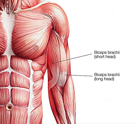
 

ANATOMY

The biceps has two tendons that attach the muscle to the bone in the shoulder and one tendon that attaches at the elbow. The tendon at the elbow is also called the distal biceps tendon. It attaches to the radial tuberosity. This is a small bump on one of the bones in your forearm (radius) near your elbow joint.
 

HISTORY AND PHYSICAL EXAMINATION

Patients with biceps tendon ruptures often present with a history of acute onset of pain in the front side of the elbow during lifting. The typical patient is a 50 years old but may be between 21 and 70 years of age; a majority of cases occur in weight lifters.

Smoking. Nicotine use can affect nutrition in the tendon.
Corticosteroid medications. Using corticosteroids has been linked to increased muscle and tendon weakness.

Patient report ecchymosis anterior to the elbow and a period of limited elbow motion secondary to acute inflammation and swelling in the region. If care is sought after the acute inflammation has , patient may present with complaints of weak elbow flexion and supination.


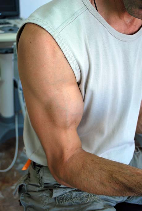
Distal biceps tendon rupture.  The tendon is contracted proximally.


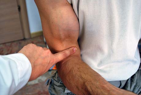
Distal biceps tendon rupture.  The Orthopaedist index finger shows the gap produced by rupture.

Long head biceps tendon rupture in young man

DIAGNOSTIC IMAGINE

Radiograph are typically normal. On occasion avulsion of the tuberosity have been demonstrated in the acute setting.
If the physical examination is conclusive, further imaging is not required.

Magnetic resonance imaging (MRI), can help to confirm the diagnosis if the physical examination findings are unclear.

Ultrasonography has also been shown to be an inexpensive and reliable method to confirm diagnosis of biceps rupture, although this technique requires a skilled ultrasonographer for accurate interpretation.


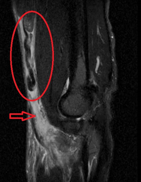
Magnetic resonance imaging: Massive rupture of distal biceps tendon, Red circle ruptured biceps tendon contracted proximaly.  Red arrow, the gap.

CONSERVATIVE TREATMENT
THERAPY: 

 CONSERVATIVE TREATMENT

Conservative treatment may be considered for patients who are elderly and inactive, or who have medical problems that make them high-risk for modest surgery.
Patients must weigh the decision to proceed with nonsurgical treatment carefully, because restoring arm function with later surgery may not be possible.

Treatment of partial rupture initially consists of a trial of nonoperative management. Treatment modalities include anti-inflammatories, splinting, and physical therapy. Although formal outcomes of nonoperative management have not been reported.

Treatment of complete tear of the biceps tendon is primarily surgical.

OPERATIVE TREATMENT
THERAPY: 

OPERATIVE TREATMENT

Treatment of complete tear in athletes is primarily surgical and this is the best way to restore both flexion and supination strength. Surgical repair is recommended in all acute cases unless advanced age or poor medical condition circumscribes operative treatment.

The surgery is done with the help of surgical microscope and microsurgical techniques for maximum patient safety and achieve better results. The duration of surgery is approximately 60-70 minutes. The patient after surgery, remains in hospital for 24 hours to obtain the maximum medical and nursing care.


Diagramatic skin incision

Distal biceps tendon rupture.  Preoperative test.


Open operative procedure, with Bicepsbutton and Biotenodesis screw (Arthrex).


Biceps tendon (green arrow), Biotenodesis screw (blue arrow)

Shematic diagram for the suturing of the biceps tendon into the bone of radius

Biceps tendon rupture pre op


Postop

THE ADVANTAGES OF THIS METHOD IS:

• the use of surgical microscope provides precise surgical manipulations and patient safety
• relatively quick mobilization of joints
• it is not necessary to apply a special brace
• short stay in the clinic
• quick return of the person in daily activities
 

POST OPERATION

Place the patient in a soft dressing or brace and allow active supination and pronation of the forearm.
Active flexion and extension as tolerated is permitted, but patients should lift nothing heavier than a coffee cup for the first four weeks.
At 6 weeks, progressive resistance exercises begin, with the goal of release to full activity by 3 months.

REHABILITATION: 

Place the patient in a soft dressing or brace and allow active supination and pronation of the forearm.
Active flexion and extension as tolerated is permitted, but patients should lift nothing heavier than a coffee cup for the first four weeks.
At 6 weeks, progressive resistance exercises begin, with the goal of release to full activity by 3 months.

Arthro Heal Clinic, www.arthrohealclinic.gr link image