LUMBAR DISC HERNIATION

INTERVERTEBRAL DISC HERNIATION

The lifetime incidence of low back pain ranges from 50-80%, whereas the incidence of sciatica ranges from 13-40%


INTERVERTEBRAL DISC ANATOMY

Intervertebral discs are found between each vertebra.
The human disc has two basic parts: an inner jell o-like center called the Nucleus Pulposus and the Annulus Fibrosis

The Nucleus Pulposus is the water-rich, gelatinous center of the disc. It has three main functions:

  • to bear or carry the downward weight (i.e., axial load) of the human body
  • act as a 'pivot point' from which all movement of the lower trunk occurs.
  • act as a ligament and bind the vertebrae together.

Lumbar spine anatomy

The Annulus Fibrosus is much more fibrous (tougher) than the nucleus. It also has a much higher collagen content and lower water content when compared to the nucleus.
Its main job is hold in place or contain the highly pressurized nucleus (the nucleus is pressurized for hold up the weight of the body), which is constantly trying to escape its central prison.

The annulus is made of concentric sheets of collagen (a tough cartilage-like substance) that are called Lamellae. The lamellae are arranged in a special configuration that makes them extremely strong and assists in their job of containing that pressurized nucleus pulposus.
 

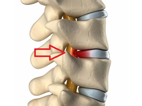
WHAT IS DISC HERNIATION?

Disc herniation occurs when the annulus fibrous breaks open or cracks, allowing the nucleus pulposus to escape. This is called a Herniated Nucleus Pulposus (HNP) or herniated disc.

  
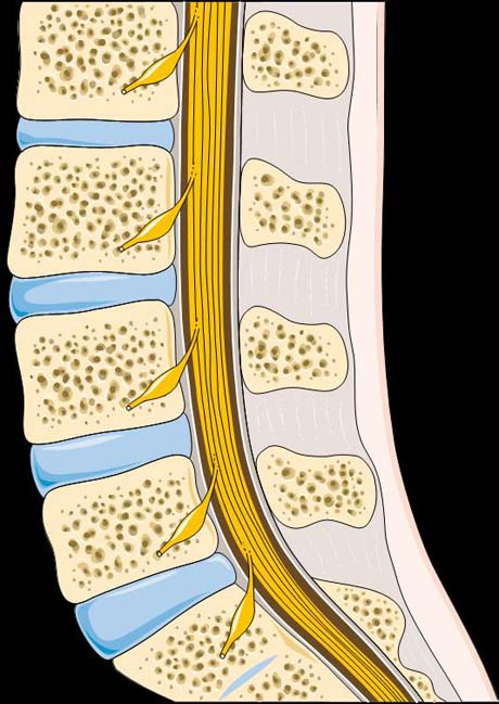
Νormal anatomy of the lumbur spine (profile)


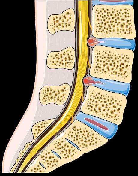
Discs herniations in two levels.  The disc protrude in to the spinal canal (red spots)
 

HISTORY OF PATIENTS WITH DISC HERNIATION

Most people relate back and leg pain to a traumatic event, but close questioning sometimes reveals that patient have had intermittent episodes of back pain for months or years.
The pain may be brought on by heavy exertion, repetitive bending, twisting, or heavy lifting. Pain usually begins in the low back and radiates to the buttock area.

Radicular pain usually extends below the knee and follows the dermatome of the involved nerve root. When the patient have sustained a disc herniation, the pain usually increases with activities, especially with sitting position.

RISK FACTORS

  • As normal consequence of aging, Intervertebral discs undergo degenerative changes, marked by dehydration of the nucleus pulposus. Progressive fibrillation, separation and tearing of annular fibers from prolonged axial and shear stress may result in a complete radial annular tear.
  •  
  • Weight lifting with improper way. Using the back muscles to lift objects, instead of the legs
  • Heavy Body weight. Exercise program create strong muscles around the lumbar spine, very important issue for healthy non herniated spine. The overweight body puts added stress on the Intervertebral discs.
  •  
  • Prolonged sitting position with wrong way. The sitting position is the worse position for the low back. The proper support during sitting with McKenzie pillow is a good option.
CONSERVATIVE TREATMENT
THERAPY: 

CONSERVATIVE TREATMENT

The treatment for vast majority of patients with a herniated disc does not normally include surgery. Eighty percent of patients will respond to conservative therapy. The primary element of conservative treatment is controlled physical activity.

THE NONSURGICAL TREATMENT INCLUDE:

Bed rest. A brief period of bed rest for 48-72 hours is beneficial for the lumbar disc herniation.

Medication. The acute phase is the proper time to use medications. The medication is started as soon as possible until the symptoms arises. Anti-inflammatory drugs oral or intramuscular injected is very useful to treat the acute low back pain.
Modern medicine exists specialized on pain due to neural inflammation exists in Orthopaedic armamentarium with very low rate of side effects as compare with classic anti-inflammatory drugs.

Physical Therapy. Physical therapy is an important treatment option for most back pain sufferers. The main ‘tool’ of physical therapy is the exercises. The exercise is for muscle strengthening and muscle stretching. Special techniques as like Mc Kenzie for mechanical evaluation and treatment of low back pain provide great advantage in therapy.
Physical therapist additionally use heat, cold, TENS (transcutaneous electrical nerve stimulation), diathermy, ultrasound, electrotherapy, and special devices for stretching the lumbar spine to relief the symptoms produced by disc herniation.
It is important that physical therapy is coupled with education, so that patients can be empowered to take charge of their own recovery.


PREVENTION

• Exercise. Regular exercise slows aging-related degeneration of the disks, and core-muscle strengthening helps stabilize and support the spine. Check with your doctor before resuming high-impact activities such as jogging, tennis and high-impact aerobics.

• Maintain good posture. Good posture reduces the pressure on your spine and disks. Keep your back straight and aligned, particularly when sitting for longer periods. Also, lift heavy objects properly, making your legs — not your back — do most of the work.

• Maintain a healthy weight. Excess weight puts more pressure on the spine and disks, making them more susceptible to herniation.

• Quit smoking. Smoking increase your risk of back problems.

  • Use the knee not your back to lift the weight.

Left: Right sitting position.    The pillow on the back support the support the whole spine (including the cervical spine)
Right:  Wrong sitting position. The deformity of the spine is obvious

OPERATIVE TREATMENT
THERAPY: 

OPERATIVE TREATMENT

The goal to surgery are neural decompression and relief of symptoms without harm to surroundings tissues. Proper patient selection is the most important factor in achieving this aims. Disc removal , intended to relieve sciatica, does not restore the normal mechanics of the lumbar spine and should not be undertaken to relieve isolated back pain without evidence of radiculopathy.

Open discektomy through a limited laminectomy and microdiskectomy remain the gold standard procedures; 90% of properly selected patient experience successful short-terms relief of symptoms. Long-term follow-up of these patient, however, reveals a progressive decline in success resulting from subsequent degenerative changes. This emphasizes the importance of postsurgical rehabilitation and proper lifelong back care by the patient.

In recent years, a trend toward the application of minimally invasive techniques has evolved, including percutaneous suction diskectomy, percutaneous laser diskectomy, percutaneous arthroscopic disc decompression and microendoscopic diskectomy.

Arthro Heal Clinic, www.arthrohealclinic.gr link image