STERNOCLAVICULAR JOINT DISLOCATION (CHRONIC)
A CASE REPORT
The dislocation of the sternoclavicular joint is a rare injury of the musculoskeletal system and usually occurs during a traffic accident or during contact sports. Generally considered high energy injuries that must be brought great powers in the anatomical region of the sternum.
The patient described was 40 years old, motorcycle driver and collided with a passing car (traffic accident). Among other fractures suffered in the lower limb had closed injury to the shoulder girdle resulting to present dislocation of the sternoclavicular joint. The sternoclavicular joint is composed of the clacicle bone and the sternum. In this position, the bones are held together by strong ligaments.
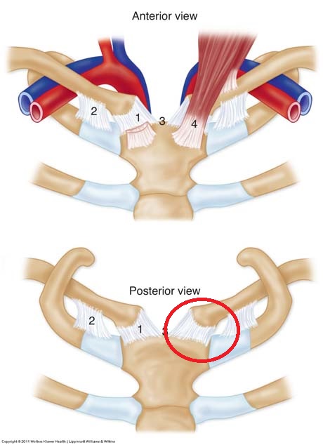
Anatomy of the sternoclavicular joint. The articulation is supported by strong ligaments which hold the bones together (clavicle - sternum), and muscle groups
When transporting the patient to the hospital initially treated fractures in the legs and not the severity of the sternoclavicular joint dislocation initially evaluated.
After six months from the traffic accident the patient has swallowing problems and slight breathing difficulties during sleep in certain postures.
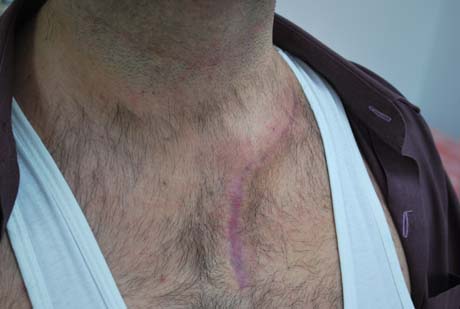
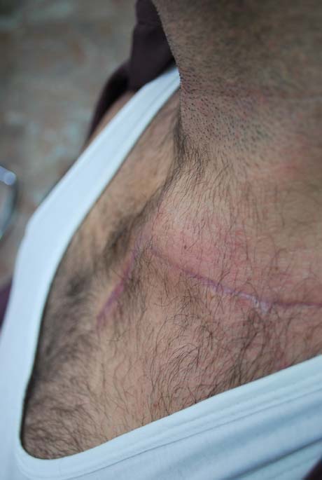
During movement of the shoulder inwardly (horizontal adduction), observe the bulge in the neck region creating the sternal end of the clavicle - moving - which creates several problems to the patient specific bedtime
Preop

Preoperative X-ray: the left sternoclavicular joint is dislocated and the clavicle has been shifted upwardly in relation the normal right clavicle
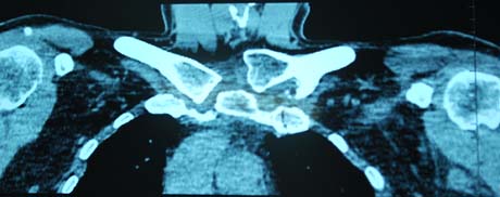
CT-scan
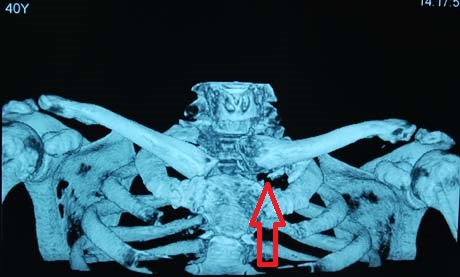
Pre op CT-scan with 3D reconstruction. The left clavicle is dislocated upward (arrow)
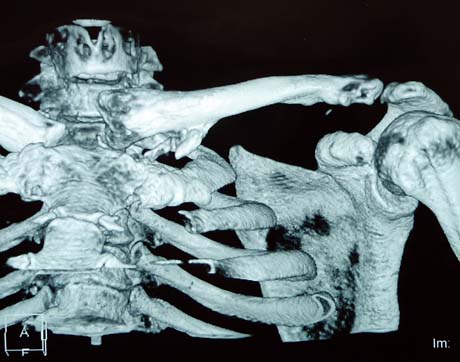
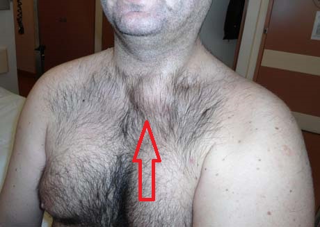
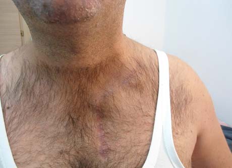
Photo showing the protrusion the sternal end of the clavicle which often penetrates the tracheal space (breathing tube ), and creates difficulty in breathing
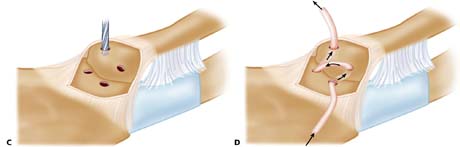
The stabilization of the the clavicle to the sternum is performed using autograft (semitendinosus muscle) which is obtained from the area of the patient's knee.
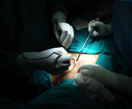
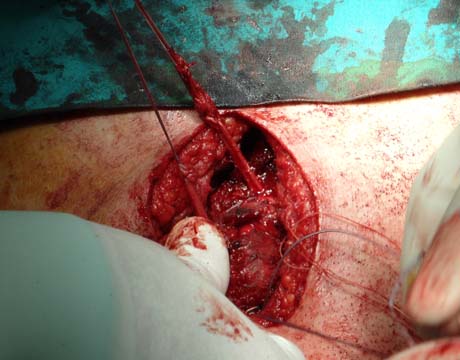
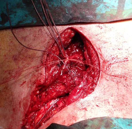
The use of autograft (semitendinosus tendon), fixes the clavicle with sternum together. It used special suture to enhance stabilization
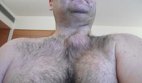
Postoperative patient profile. The distortion is completely corrected.
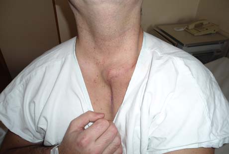
The projection of the sternal end of the clavicle is evident.
Because the surgery in which the patient was failed to resolve the problem is decided to second surgery.

During the second surgery was done to remove the sternal end of the clavicle and the rest of the clavicle stabilized to the first rib.
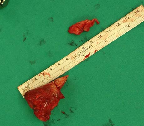
Remove about 2.5 cm of the sternal end of the clavicle
Two months after the second surgery. The aesthetic and functional assessed by the patient himself as great. There are no functional problems regarding breathing and the patient now can be progressively return to professional activities
The patient is evaluated a regular basis for three years after the second surgery and showed no sensory or motor problem in the injury of the clavicle area. He lives a normal life and working normally. No mention pain during effecting the movements of the left arm.